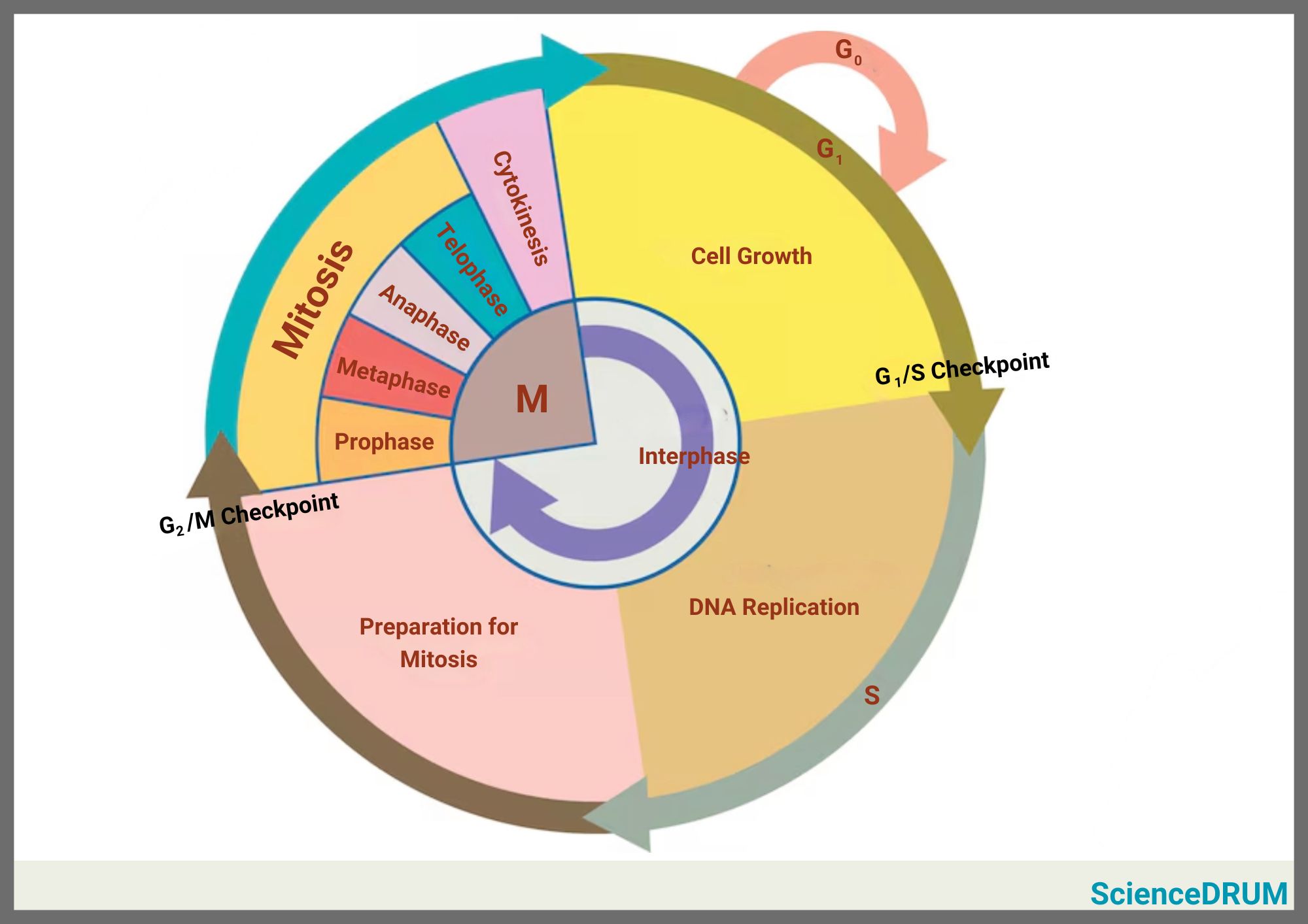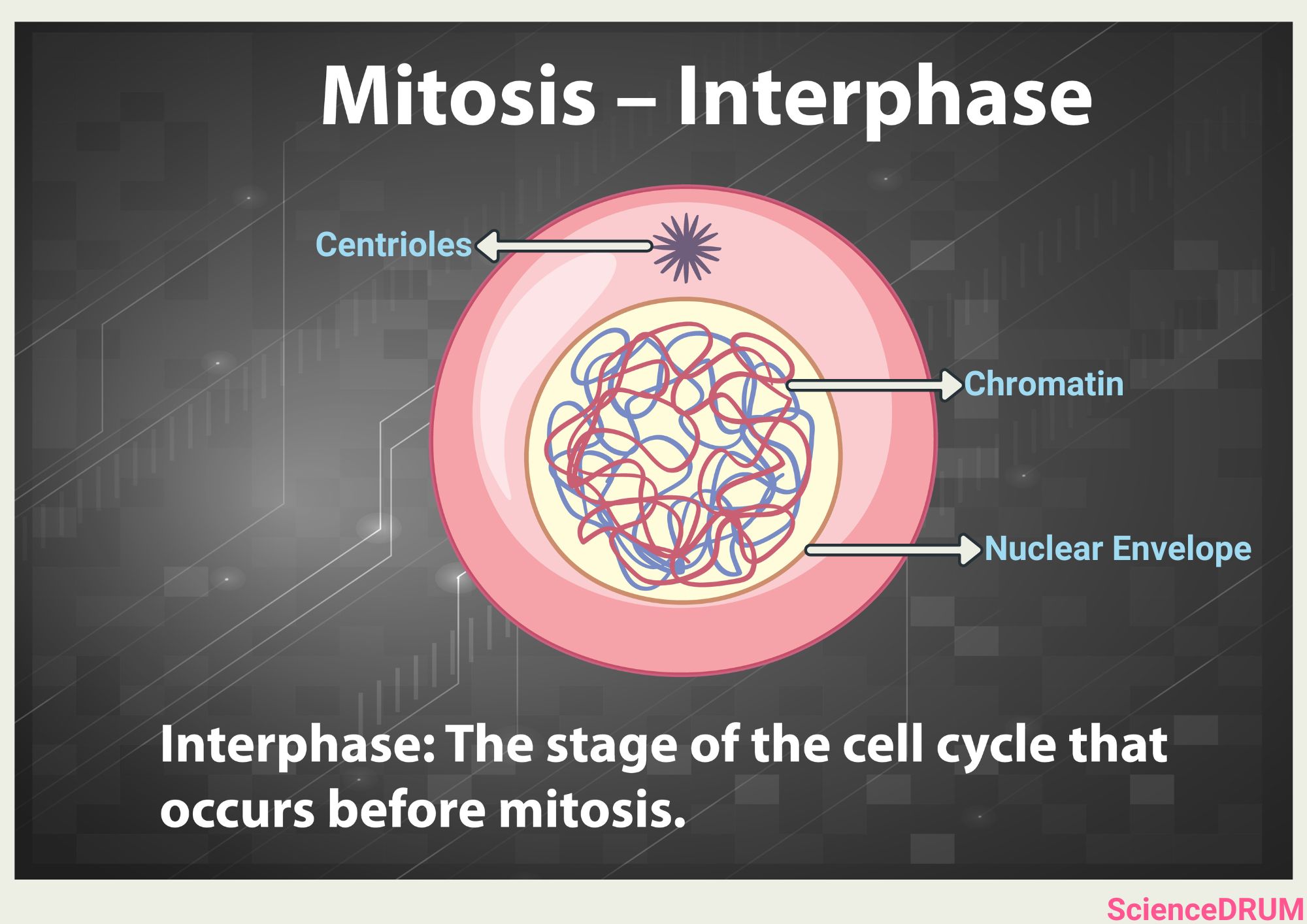
A few reasons that make it difficult to observe individual chromosomes under a light microscope during interphase are:
- During interphase, chromosomes are highly extended and diffused as compared to mitosis, making it difficult to identify them.
- The size of the chromosomes during interphase is typically below the resolving power of a light microscope, making it difficult to observe them.
- The nucleus is crowded and complex and contains many other structures such as the nucleolus, nuclear envelope, and cytoplasmic extensions, all of which obscure the view of individual chromosomes.
- Chromosomes also do not have the characteristic “X” shape during interphase, making it harder to distinguish them from other structures in the nucleus.
This article explains how light microscopy is used to observe chromosomes, why it’s not the best option to study chromosomes during interphase, and some of the most widely used alternative techniques to observe interphase chromosomes.
How Light Microscopy Works?
Light microscopy is the most commonly used method for observing cells and their structures. The basic principle of light microscopy involves passing light through a sample and using lenses to magnify the image.
However, there are limitations to what can be observed using light microscopy. The resolution of light microscopy is limited to approximately 200 nanometers. This means that structures smaller than 200 nanometers cannot be distinguished from each other.
Why Is it Difficult to Observe Individual Chromosomes With a Light Microscope During Interphase?

In general, the resolving power of a microscope is determined by the wavelength of the light used to illuminate the sample. Since visible light has a wavelength of about 400-700 nanometers, this sets the limit of how small an object can be resolved by a light microscope.
- Chromosomes during interphase are much more extended and diffused than during mitosis and their size is usually below the resolving power of a light microscope, which makes them difficult to observe.
- Furthermore, the nucleus is a crowded and complex environment with many other structures, such as the nucleolus, nuclear envelope, and cytoplasmic extensions, that can obscure the view of individual chromosomes.
- Interphase chromosomes also do not have the characteristic “X” shape that they have during mitosis, making them harder to distinguish from other structures in the nucleus.
- Another factor that makes it difficult to observe interphase chromosomes with a light microscope is their degree of condensation. Interphase chromosomes exist in a less compact state than those during mitosis, where they are highly condensed and can be detected easily. The level of chromosome condensation varies between different regions of the chromosome, and even within the same chromosome during different stages of the cell cycle.
These are some of the main reasons why chromosomes are not visible under the light microscope.
Alternative Techniques for Observing Interphase Chromosomes
One of the biggest challenges in studying interphase chromosomes is their thin and thread-like structure, which makes them difficult to see with a light microscope. But recent advancements in technology have made it possible to develop multiple tools to observe interphase chromosomes in more detail.
One such technique is electron microscopy. Unlike light microscopy, which uses visible light to magnify structures, electron microscopy uses beams of electrons. This technique can provide much higher magnification and resolution, allowing scientists to see individual chromosomes and their components in greater detail. {1}
There are two main types of electron microscopy:
- Transmission electron microscopy (TEM). In TEM, a beam of electrons is transmitted through a thin sample, and the resulting image is formed by the electrons that pass through the sample. This technique can provide very high-resolution images of chromosomes and their components. However, preparing the sample for TEM can be challenging, and the process can damage the sample.
- Scanning electron microscopy (SEM). In SEM, a beam of electrons is directed onto the surface of a sample, and the resulting image is formed by the electrons that bounce off the surface. This technique can provide three-dimensional images of chromosomes and their components, allowing scientists to observe their shape and structure in more detail.
Another powerful technique used to observe interphase chromosomes is fluorescence microscopy, which involves the use of fluorescent dyes that bind to DNA molecules in the chromosomes.
When the dye is illuminated with a specific wavelength of light (400-700 nanometers), it emits a fluorescent glow that makes chromosomes visible. Fluorescence microscopy can provide high-resolution images of chromosomes and their components, and it can also be used to observe chromosomes in living cells.
This makes it a powerful tool for studying the behavior of chromosomes during interphase. There are several types of fluorescence microscopy including confocal microscopy and super-resolution microscopy.
- Confocal microscopy. This technique uses a laser to illuminate a single point on the sample, and the resulting image is formed by detecting the fluorescent light emitted from that point. This technique can provide high-resolution images of chromosomes and their components, and it can also be used to create three-dimensional images of the sample.
- Super-resolution microscopy. Also known as nanoscopy, this technique is relatively new and allows scientists to observe structures that are smaller than the diffraction limit of light. This technique uses advanced imaging methods to bypass the diffraction limit of light and provide very high-resolution images of chromosomes and their components. Super-resolution microscopy has revolutionized the field of microscopy and has allowed scientists to observe interphase chromosomes in unprecedented detail. {2} {3}
In addition to these techniques, there are also specialized microscopy methods that are used to study specific aspects of interphase chromosomes. For example, fluorescence in situ hybridization (FISH) is a technique that is used to detect specific DNA sequences in chromosomes. {4}
FISH is also used to photograph chromosomes and prepare karyotypes. It involves labeling a specific DNA sequence with a fluorescent dye and then observing its location within the chromosome.
Another technique, called chromatin immunoprecipitation (ChIP), is used to study the interactions between the the chromosomal components. {5} This technique involves cross-linking DNA and proteins in the chromosome and then using an antibody to isolate the DNA-protein complex.
The isolated DNA can then be sequenced to determine which proteins are bound to specific regions of the chromosome.
Observing interphase chromosomes is essential to understand how genes are expressed and how they contribute to specific traits and characteristics. Although light microscopy has its limitations, scientists have developed alternative techniques and advanced microscopy technologies that allow us to observe chromosomes in unprecedented detail.
Observing Interphase Chromosomes
Observing individual chromosomes during interphase is difficult because of their small size, their relatively diffuse and extended structure, and the complexity of the nucleus.
While light microscopy is limited in its ability to resolve these structures, there are other techniques that can provide more detailed information about chromosome structure and behavior.
Frequently Asked Questions
Sources
1 – University of Massachusetts: “What is Electron Microscopy?”
2 – Carleton College: “What Is Fluorescent Microscopy?”
3 – Cold Spring Harbor Protocols: “Fluorescence Microscopy.”
4 – National Human Genome Research Institute: “Fluorescence In Situ Hybridization (FISH).”
5 – Cold Spring Harbor Protocols: “Chromatin Immunoprecipitation (ChIP).”