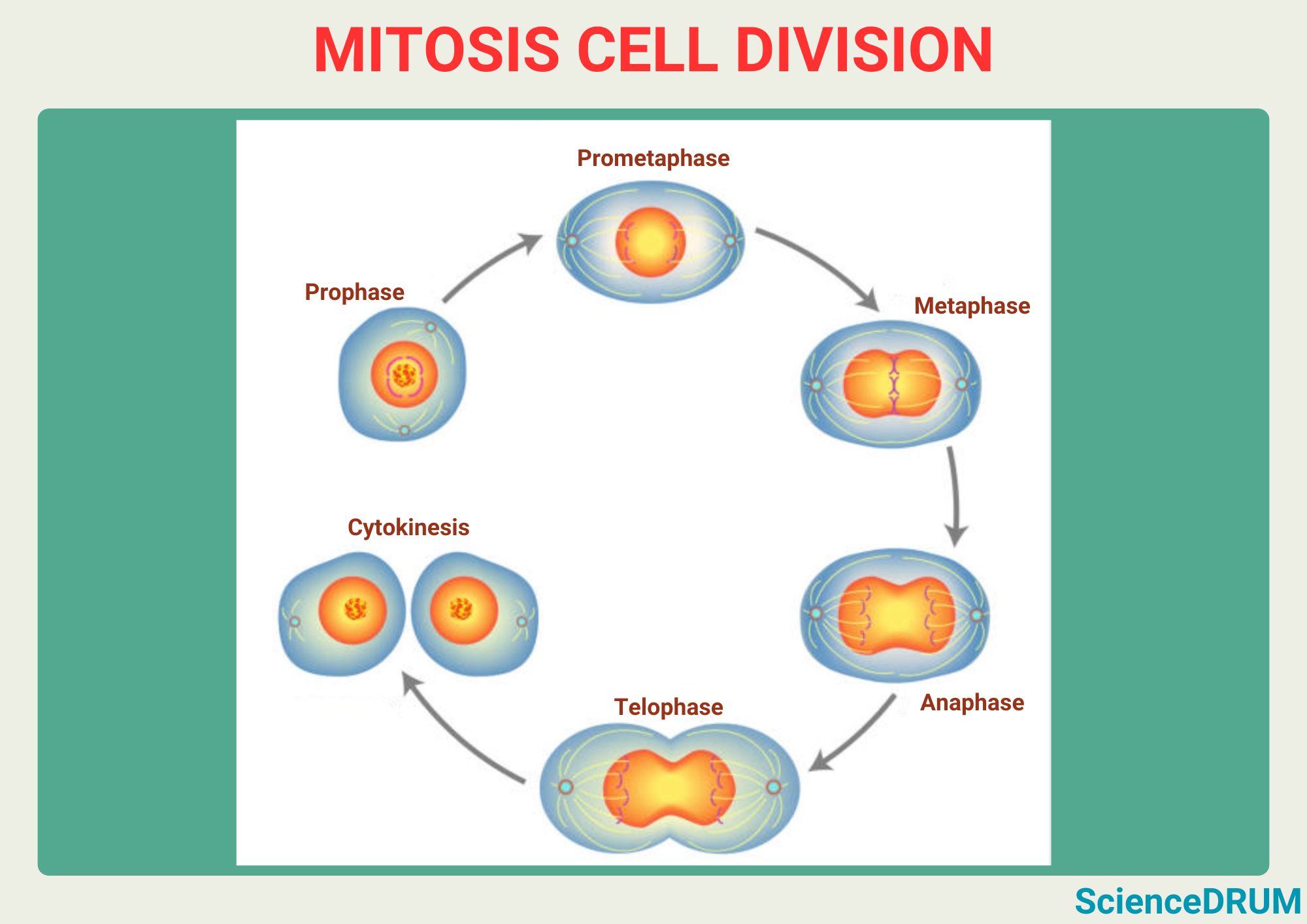
The structure that forms in prophase along which the chromosomes move is the mitotic spindle. This dynamic and complex structure is made up of microtubules that grow out from two poles located at opposite ends of the cell, and it plays a critical role in organizing the chromosomes and pulling them apart as the cell divides.
The process of spindle formation and chromosome alignment is tightly regulated and involves many proteins and pathways, and aberrations in these processes can lead to serious consequences for the health of the cell.
This article explains the structures that form during prophase and work to move chromosomes and the intricate process of mitosis that they help to orchestrate.
- Prophase and the Chromosome Structure
- What Structure Forms in Prophase Along Which the Chromosomes Move?
- Other Structures Involved in Spindle Fiber Formation and Chromosome Separation
- Formation of Kinetochore-Microtubule Attachments in Prophase
- Regulation of Spindle Assembly and Chromosome Alignment in Prophase
- Aberrations in Spindle Formation and Their Consequences
- Frequently Asked Questions
Prophase and the Chromosome Structure
Mitosis is the process by which a cell divides into two identical daughter cells. It is a crucial part of growth, development, and repair in multicellular organisms.
Prophase, the first of four mitosis stages, is when the genetic material in the cell, which is organized into structures called chromosomes, begins to contract and become visible under a microscope.
This condensation allows the chromosomes to be more easily separated and distributed to the two daughter cells. Chromosomes are made up of DNA and protein.
The DNA contains the genetic information that is passed from parent to offspring, while the proteins help organize and protect the DNA. Chromosomes are organized into pairs, with one chromosome in each pair coming from each parent. Humans have 23 pairs of chromosomes, for a total of 46 chromosomes in each cell.
What Structure Forms in Prophase Along Which the Chromosomes Move?
During prophase, a structure called the spindle apparatus (also called the mitotic spindle) begins to form. The spindle apparatus is made up of spindle fibers, which are long protein strands that stretch from opposite poles of the cell.
The spindle fibers attach to the chromosomes via a protein complex called the kinetochore. The spindle fibers then pull the chromosomes towards the center of the cell.
Other Structures Involved in Spindle Fiber Formation and Chromosome Separation
The spindle apparatus is a dynamic structure that undergoes continuous reorganization during mitosis. During prophase, the spindle apparatus begins to form by the nucleation of microtubules from two centrosomes, which are located at opposite poles of the cell.
Role of Centrosomes in Spindle Fiber Formation
The spindle fibers are anchored to the cell’s centrosomes, which are structures located near the nucleus. The centrosomes play a critical role in organizing the spindle fibers and ensuring that they are evenly distributed to the two daughter cells.
Kinetochores: Protein Complexes that Attach to Chromosomes
The kinetochore is a protein complex that forms at the centromere of each chromosome. The spindle fibers attach to the kinetochore and use it to move the chromosomes towards the center of the cell.
Microtubules: The Building Blocks of Spindle Fibers
Spindle fibers are made up of microtubules, which are tiny protein tubes. These microtubules are dynamic structures that can grow and shrink as needed to adjust the position of the chromosomes.
The microtubules grow out from the centrosomes and extend towards the chromosomes, forming a network of microtubules called the spindle fibers and extend towards the center of the cell. As the microtubules grow, they search for and capture the chromosomes, which are now compact and can be observed under the microscope.
Microtubules act as a scaffold that helps to organize the chromosomes and pull them apart.
- Polar Microtubules. In addition to the spindle fibers that attach to the kinetochore, there are other microtubules called polar microtubules. These microtubules do not attach to the chromosomes but instead push against each other to help separate the chromosomes into two groups. {1}
- Astral Microtubules. Astral microtubules are microtubules that radiate out from the centrosomes towards the cell membrane. They help orient the spindle apparatus and ensure that the spindle fibers are aligned correctly. {2}
Molecular Motors: The Force Behind Chromosome Movement
The movement of the chromosomes is powered by molecular motors, which are proteins that use energy to move along the microtubules. These motors “walk” along the microtubules, pulling the chromosomes towards the center of the cell. {3}
The Spindle Poles: Spindle Assembly and Chromosome Segregation
The spindle poles are critical components of the spindle apparatus, as they serve as the anchoring points for the microtubules. During prophase, the two spindle poles are separated from each other, and they begin to migrate towards opposite ends of the cell.
This movement is facilitated by the motor proteins that are attached to the microtubules, which pull the poles apart.
Formation of Kinetochore-Microtubule Attachments in Prophase
During prophase, the kinetochore-microtubule attachments are formed, which are essential for chromosome segregation. The kinetochore is a complex protein structure that forms at the centromere region of the chromosome.
It acts as a platform for the attachment of the microtubules, which form a network around the chromosome. The kinetochore then uses the microtubules as a “tug of war” force to pull the chromosome towards the center of the cell.
Regulation of Spindle Assembly and Chromosome Alignment in Prophase
The assembly of the spindle apparatus and the alignment of the chromosomes are tightly regulated processes that involve many proteins and pathways. One of the key regulators of spindle assembly is the protein kinase Aurora B, which is responsible for phosphorylating many of the proteins involved in spindle formation and chromosome alignment.
Another important regulatory pathway is the spindle checkpoint, which ensures that all chromosomes are properly attached to the spindle before the cell proceeds to the next stage of mitosis. This checkpoint prevents the cell from dividing prematurely and potentially causing genomic instability.
Aberrations in Spindle Formation and Their Consequences
Spindle formation and chromosome alignment are complex processes that can go wrong in many ways. For example, if the spindle apparatus fails to properly capture and pull apart the chromosomes, it can lead to chromosome segregation errors that result in aneuploidy.
This can cause developmental defects and can also contribute to cancer progression. Additionally, defects in spindle formation can lead to mitotic arrest or apoptosis, which can have serious consequences for the health of the cell.
Frequently Asked Questions
Sources:
1 – Journal of Cell Biology: “Long astral microtubules uncouple mitotic spindles from the cytokinetic furrow.”
2 – Cell Cycle: “Astral microtubules control redistribution of dynein at the cell cortex to facilitate spindle positioning.”
3 – Alberts, B., Johnson, A., Lewis, J. “Molecular Biology of the Cell.” Garland Science, 2002.